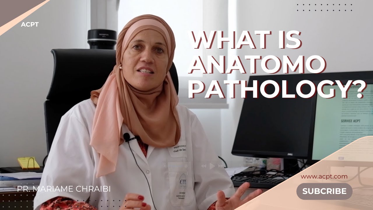
What is Anatomopathology?
An overview of the fundamental steps and techniques in anatomic pathology — from macroscopic examination to immunohistochemistry.
Macroscopic Examination (Grossing)
The first step in tissue evaluation involves the macroscopic (gross) examination of surgical specimens.
- Describing the specimen's size, shape, color, and consistency
- Identifying lesions or abnormal areas
- Selecting representative sections for microscopic analysis
Tissue Processing
To prepare tissue for microscopic examination, it must undergo several processing steps.
Preserves tissue architecture and prevents degradation. Formalin is the most commonly used fixative.
After dehydration and clearing, tissues are infiltrated with paraffin wax to maintain structure.
Thin sections (typically 3–5 µm) are cut using a microtome and placed on glass slides.
Stains such as hematoxylin and eosin (H&E) enhance contrast, allowing visualization of cell and tissue morphology.
Introduction to Immunohistochemistry (IHC)
IHC is a powerful technique that uses antibodies to detect specific antigens in tissues. It plays a critical role in:
- Differentiating tumor types
- Identifying prognostic and predictive markers
- Guiding targeted therapy
Frequently Asked Questions
Anatomic pathology examines tissue architecture through biopsies and surgical specimens, while cytology focuses on the examination of individual cells, typically from fluid samples, smears, or fine-needle aspirates.
Formalin fixation halts tissue autolysis and preserves cellular and structural detail, ensuring the tissue remains representative of its living state throughout processing and staining.
IHC is used when routine H&E staining is insufficient to characterize a lesion — for example, to confirm tumor origin, identify specific biomarkers, or guide the selection of targeted therapies.
More Video Training
We're preparing additional training videos covering more topics in anatomic pathology. Check back soon.
Have a question about this topic?
Our team is available to help with any technical questions or training needs related to anatomic pathology.
Contact our team